Avisena Specialist Hospital Logo
Appointments +(603) 5515 1966
General Line +(603) 5515 1888
Highlights / Articles

Articles

June 27, 2025

Fractures in Focus: Classification, Risk Factors, Diagnosis, and Modern Management

A fracture, commonly known as a broken bone, is a disruption in the continuity of bone tissue. From a minor hairline crack to a complete shattering of bone, fractures can affect anyone irrespective of age.

While often associated with sudden traumatic events like falls or accidents, they can also result from repetitive stress or underlying medical conditions that weaken bone structure.

Understanding fractures – how they are classified, what factors increase their risk, how they are accurately diagnosed, and the array of modern management strategies available – is crucial for healthcare professionals and public awareness and prevention. This article delves into these key aspects, providing a comprehensive overview.


What is a Fracture?

At its core, a bone fracture is a break. Bones are remarkably strong and resilient, designed to withstand considerable force. However, when the force applied to a bone exceeds its structural capacity, it breaks. This force can be a sudden, high-impact trauma (e.g., a car accident, a fall from height), or it can be a lesser force applied repeatedly over time (stress fracture), or even minimal force if the bone is already compromised by disease (pathological fracture). The process of bone healing is a complex biological cascade, involving inflammation, soft callus formation, hard callus formation, and eventual bone remodelling, aiming to restore the bone to its original strength and shape.


Classification of Fractures

Fractures are classified in numerous ways, providing a systematic approach for diagnosis, communication among clinicians, and guiding treatment strategies. Key classification criteria include:

Based on Skin Integrity (Open vs. Closed)

Fractures may be of two types based on skin integrity:

  • Closed (Simple) Fracture: The skin overlying the fractured bone remains intact. While the bone is broken internally, there is no open wound connecting the fracture site to the external environment. This type generally carries a lower risk of infection.
  • Open (Compound) Fracture: The skin and soft tissues are disrupted, exposing the fractured bone to the outside environment. This can occur either because the bone fragments pierce the skin, or the trauma itself creates a wound. Open fractures are considered medical emergencies due to the high risk of infection and the potential for significant soft tissue damage.

Based on Fracture Pattern/Morphology

Fractures may be classified by the way the bone has broken:

  • Transverse Fracture: The break occurs in a straight line, perpendicular to the long axis of the bone. Often results from a direct blow.
  • Oblique Fracture: The break occurs at an angle across the bone, usually due to a twisting force.
  • Spiral Fracture: The fracture line spirals around the bone, typically caused by a twisting or rotational injury.
  • Comminuted Fracture: The bone is broken into three or more fragments. This indicates high-energy trauma and can be more complex to treat.
  • Segmental Fracture: A rare type of comminuted fracture where a segment of the bone is isolated from the main parts.
  • Impacted Fracture: One end of the bone fragment is driven into the other, often seen in falls where bones are “telescoped” into each other.
  • Greenstick Fracture: An incomplete fracture common in children, where one side of the bone is broken and the other is bent (like a green stick). Children’s bones are more flexible.
  • Torus (Buckle) Fracture: Also common in children, where the bone buckles or bulges on one side, without a complete break.
  • Avulsion Fracture: A small piece of bone is pulled away from the main bone by a strong ligament or tendon.
  • Compression Fracture: Occurs when a bone is crushed, particularly common in the vertebrae due to osteoporosis or severe trauma.

Fractures based on Displacement

Fractures may be of two types based on displacement:

  • Non-displaced Fracture: The bone breaks, but the fragments remain in their proper alignment.
  • Displaced Fracture: The bone fragments have moved out of their normal alignment, requiring reduction (realignment) before immobilisation.

Fractures based on Location

Fractures are also classified by the location of the specific bone (e.g., femoral fracture, radial fracture) and its part (e.g., neck of femur fracture, distal radius fracture).


Other Specific Types of Fractures

Fractures can be of specific types such as:

  • Stress Fracture: A tiny crack in a bone caused by repetitive stress, rather than a single forceful injury. Common in athletes (e.g., shin splints, foot fractures).
  • Pathological Fracture: A fracture that occurs in a bone already weakened by an underlying disease, such as osteoporosis, bone tumours (primary or metastatic), or osteomyelitis. The force involved might be minimal, or even no trauma at all.
  • Epiphyseal (Growth Plate) Fracture: Fractures that occur through the growth plate (physis) in children. These require careful management as they can affect future bone growth. Classified using the Salter-Harris system.

Risk Factors for Fractures

While accidents can happen to anyone, certain factors significantly increase an individual’s susceptibility to fractures:

Age

  • Children: More prone to greenstick and torus fractures due to bone flexibility, and growth plate injuries.
  • Elderly: Increased risk of fractures due to osteoporosis, falls, and slower reaction times. Hip, wrist, and vertebral fractures are common.

Bone Density (Osteoporosis)

  • This is the most significant risk factor for fractures in older adults, particularly post-menopausal women. Osteoporosis leads to weakened, brittle bones that can fracture easily, even from minor falls or everyday activities.

Nutritional Deficiencies

  • Calcium and Vitamin D: Insufficient intake of these crucial nutrients impairs bone mineralisation and strength. Vitamin D is essential for calcium absorption.
  • Malnutrition: General poor nutrition can weaken bones and muscles, increasing fall risk.

Lifestyle Factors

  • Physical Inactivity: Lack of weight-bearing exercise weakens bones.
  • Excessive Alcohol Consumption: Can lead to poor nutrition, increased fall risk, and reduced bone density.
  • Smoking: Reduces bone density and impairs bone healing.
  • Eating Disorders: Anorexia nervosa can lead to severe bone loss.

Medical Conditions

  • Endocrine Disorders: Hyperthyroidism, hyperparathyroidism, Cushing’s syndrome, diabetes.
  • Gastrointestinal Disorders: Celiac disease, inflammatory bowel disease (IBD) that affects nutrient absorption.
  • Rheumatological Conditions: Rheumatoid arthritis, which can cause bone erosion is often treated with corticosteroids that weaken bones.
  • Kidney Disease: Can affect calcium and phosphate balance.
  • Certain Cancers: Multiple myeloma, bone metastases.
  • Neurological Conditions: Parkinson’s disease, stroke, epilepsy, which increase fall risk.

Medications

  • Long-term use of corticosteroids, some anti-seizure medications, certain cancer treatments, and proton pump inhibitors can increase fracture risk.

Environmental Factors

  • Poor lighting, slippery surfaces, cluttered homes, and lack of assistive devices can increase fall risk, especially in the elderly.

Diagnosis of Fractures

Accurate and timely diagnosis is critical for effective fracture management. The process typically involves:

Clinical Assessment

Diagnosis typically begins with a clinical assessment. This involves history taking, where healthcare professionals gather details about the injury’s mechanism (like how a fall happened), the onset of pain, any pre-existing conditions, and current medications. This is followed by a thorough physical examination, which includes carefully checking for swelling, bruising, deformity, and tenderness over the bone, as well as assessing the range of motion (though this might be limited by pain). Crucially, a neurovascular assessment is performed to check pulses, sensation, and motor function distal to the injury, vital for ruling out any nerve or blood vessel damage.

Imaging Studies

Imaging studies may be of various types as elucidated below:

  • X-rays: These are the primary diagnostic tools. Multiple views (at least two, usually perpendicular) are taken to visualise the fracture line, displacement, and bone alignment. X-rays are excellent for detecting bone breaks but can miss stress fractures or subtle soft tissue injuries.
  • CT (Computed Tomography) Scan: Provides detailed cross-sectional images of bone, offering superior clarity for complex fractures (e.g., pelvis, spine, joints), assessing comminution, and pre-operative planning. It’s also useful for identifying occult (hidden) fractures not visible on X-rays.
  • MRI (Magnetic Resonance Imaging): Excellent for visualising soft tissues (ligaments, tendons, cartilage, muscles) as well as bone marrow oedema, making it ideal for detecting stress fractures, occult fractures, bone bruising, and soft tissue injuries associated with fractures.
  • Bone Scan (Nuclear Medicine Scan): Can detect increased bone turnover, useful for diagnosing stress fractures, occult fractures, or identifying bone tumours and infections, particularly when other imaging is inconclusive.

Modern Management of Fractures

Fracture management aims to achieve bone healing, restore function, alleviate pain, and prevent complications. Treatment approaches have become increasingly sophisticated, moving towards more stable fixation and earlier rehabilitation.

The “R” Principles of Fracture Management

The R principles of fracture management involve the following steps:

Reduction

Realigning the displaced bone fragments. This can be:

  • Closed Reduction: Manipulating the bone fragments externally without surgical incision.
  • Open Reduction: Surgical intervention is required to expose the fracture site and directly realign the fragments.

Retention/Immobilisation

Maintaining the reduced fragments in proper alignment until healing occurs. Methods include:

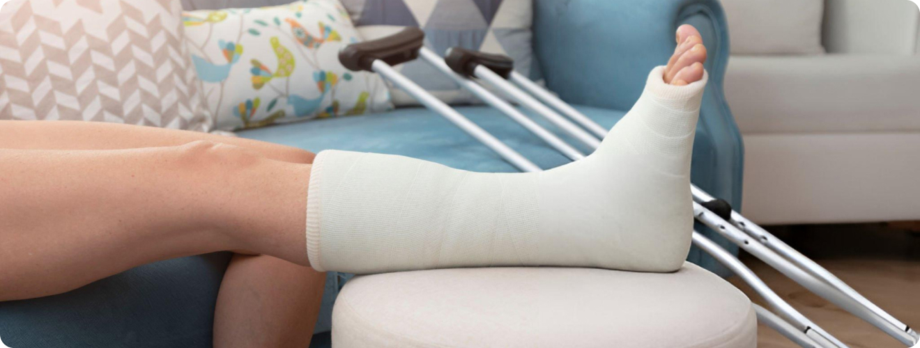
  • Casts and Splints: External immobilisation for stable fractures, protecting the bone while it heals.
  • Traction: Using weights to apply a pulling force to realign bones, is less common now due to surgical advancements.
  • External Fixation: Pins are inserted into the bone fragments and connected to an external frame outside the body. Used for complex open fractures, highly comminuted fractures, or when internal fixation is temporarily not feasible.
  • Internal Fixation (ORIF – Open Reduction Internal Fixation): Surgical insertion of metal implants (plates, screws, rods/nails, wires) directly into the bone to hold fragments together. This provides rigid stability, allowing for earlier movement and rehabilitation.

Non-Surgical Management (Conservative)

Non-surgical management is suitable for non-displaced or minimally displaced fractures and those that are inherently stable. This approach primarily involves immobilisation using casts, splints, or braces, alongside pain management with analgesics. Regular follow-up X-rays are crucial to monitor healing progress. Once the initial immobilisation is removed, rehabilitation through physiotherapy becomes vital to help patients regain strength, range of motion, and overall function.

Surgical Management

Surgical management is followed for displaced, unstable, or open fractures, those involving joints, or cases of non-union (failure to heal) or malunion (incorrect healing). Common surgical techniques include Open Reduction Internal Fixation (ORIF), where bone fragments are directly realigned and secured with implants, offering precise reduction. For long bones, Intramedullary Nailing is highly effective, involving the insertion of a metal rod into the bone’s hollow centre for stabilisation. In severe joint fractures, particularly in older adults, Arthroplasty (Joint Replacement) may be performed to replace part or all of the damaged joint.

Modern Advancements in Fracture Care

There have been significant advancements in fracture care such as:

  • Minimally Invasive Surgery: Techniques using smaller incisions and specialised instruments (e.g., percutaneous plating, intramedullary nailing without extensive exposure) to reduce soft tissue damage, pain, and recovery time.
  • Biologics and Bone Stimulators: Research into enhancing bone healing using growth factors, bone morphogenetic proteins (BMPs), platelet-rich plasma (PRP), and electrical/ultrasound bone stimulators. These are typically used for difficult-to-heal fractures (non-unions).
  • Improved Implant Technology: Stronger, more biocompatible materials and anatomically contoured plates and screws that provide better fixation and stability.
  • Advanced Imaging: Real-time fluoroscopy during surgery for precise implant placement, and 3D printing for pre-operative planning of complex cases.
  • Early Mobilisation and Rehabilitation: A major shift towards getting patients moving sooner. Modern stable fixation techniques allow for earlier weight-bearing and physiotherapy, preventing stiffness, and muscle wasting, and promoting faster return to function. This proactive rehabilitation is critical for optimal outcomes.
  • Telemedicine and Remote Monitoring: Increasingly used for follow-up appointments and monitoring progress, improving patient convenience and access to care.

Rehabilitation

Rehabilitation is integral to fracture management, starting almost immediately after injury, even while the affected limb is immobilised, through exercises for unaffected body parts. This crucial process includes physiotherapy to regain range of motion, strength, balance, and proprioception. Occupational therapy then helps patients adapt to daily activities and regain their independence. Pain management remains an ongoing and essential component throughout this healing and recovery journey.


The Bottom Line

Fractures, though common, represent a diverse range of injuries, each demanding a tailored approach to management. From the swift diagnosis facilitated by advanced imaging technologies to sophisticated surgical techniques and comprehensive rehabilitation protocols, the field of orthopaedics has made remarkable strides in improving patient outcomes.

Understanding the various classifications of fractures, recognising their associated risk factors, and appreciating the intricacies of modern diagnosis are fundamental steps. Crucially, the emphasis now extends beyond mere bone healing to ensuring a full restoration of function, minimising long-term disability, and enhancing the overall quality of life for individuals affected. As research continues to uncover new insights into bone biology and refine treatment methodologies, the future of fracture care promises even greater precision and more effective pathways to recovery.


Frequently Asked Questions

What are the 7 types of fractures?

The 7 common types of fractures include Transverse, Oblique, Spiral, Comminuted, Greenstick, Stress, and Open (Compound).

What does fracture mean?

A fracture means a broken bone, where there’s a partial or complete break in the bone’s continuity.

What are the 10 causes of fractures?

The 10 common causes of fractures include Traumatic injuries (falls, accidents, sports), osteoporosis, nutritional deficiencies (calcium, Vitamin D), physical inactivity, excessive alcohol consumption, smoking, certain medical conditions (e.g., endocrine, GI disorders, cancer), long-term medication use (e.g., corticosteroids), repetitive stress/overuse, and environmental hazards (increasing fall risk).


Back to Articles